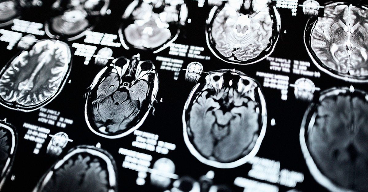
Parkinson’s disease is a progressive brain disorder that can cause shaking, stiffness, and difficulty with balance and coordination. Scientists are now exploring the use of MRI scans to predict disease progression in people with Parkinson’s. According to a study published in Radiology, MRI scans can help map the structural and functional connections in the brain, allowing researchers to predict patterns of atrophy in individuals with the disease.
The study utilized MRI data from 86 people with mild Parkinson’s disease and 60 healthy controls to map the brain’s neural connections. Researchers found that having Parkinson’s disease for one or two years correlated with atrophy at two to three years past the baseline. Additionally, the models developed by the researchers were able to predict gray matter atrophy accumulation over three years in certain parts of the brain. This suggests that the brain’s structural and functional connectome significantly influences gray matter atrophy progression in Parkinson’s disease.
Massimo Filippi, a senior author of the study, emphasized that the findings could enhance clinical trials for Parkinson’s disease and its treatments. By incorporating disease exposure indexes based on healthy brain connectivity and atrophy severity in patients, medical professionals can better predict the progression of neurodegenerative changes in Parkinson’s. This targeted approach can lead to more efficient and effective trials, accelerating the development of new therapies for the disease.
While conventional MRI scans are not used to diagnose Parkinson’s, they can help differentiate it from secondary causes of parkinsonian symptoms. The disease primarily affects individuals over the age of 60 and is more common in men than in women. Symptoms include unintended movements, stiffness, and difficulty with balance. There is currently no cure for Parkinson’s, but medications such as Levodopa, dopamine agonists, and enzyme inhibitors can help manage symptoms. Some individuals may benefit from deep brain stimulation, where electrodes are implanted in the brain to help regulate movement.
In conclusion, the use of MRI scans to predict disease progression in Parkinson’s disease shows promise in advancing our understanding of the disease and improving clinical trials. By mapping the brain’s neural connections and predicting patterns of atrophy, researchers can better predict the progression of neurodegenerative changes in Parkinson’s. This approach may lead to more efficient and effective trials, ultimately accelerating the development of new therapies for the disease. Further research in this area could have significant implications for the future of Parkinson’s treatment.


